
Unveiling the emergence of multidrug-resistant pathogens in exotic pets from France: a comprehensive study (2017-2019)
 0
0 , ...
, ... Abstract
Aim: This study intends to assess the occurrence of multi-drug resistant (MDR) resistant pathogens among exotic pets from France (2017-2019).
Methods: Isolates were identified using MALDI-TOF-MS. Antimicrobial susceptibility testing was conducted for 21 antimicrobials and was assayed by disk diffusion methods. Statistical analyses were carried out using GraphPad Prism® (version 9.4.1).
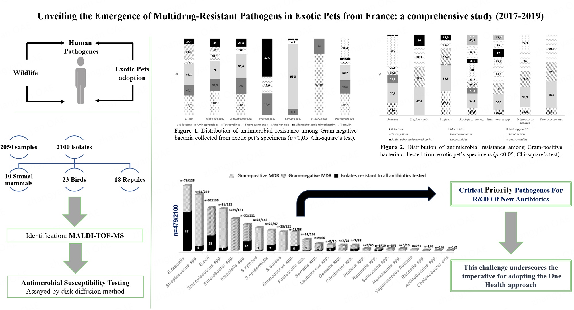
Results: Isolates (n = 2,100) recovered from samples of 10 small mammals (n = 1,555), 23 birds (n = 287), and 18 reptiles (n = 208) species were identified as Enterobacterales (n = 634), Pseudomonadaceae (n = 176), Pasteurellacea (n = 276), Staphylococcaceae (n = 563),Streptococcaceae (n = 259), and Enterococcaceae (n = 186). Consistent high resistance rates were observed among diverse genera and/or species to beta-lactams, tetracyclines, and macrolides. Notably, a significant prevalence of MDR bacteria was identified, with 22.8% (n = 479/2,100,
Conclusions: This study emphasizes exotic pets as an emergent reservoir of MDR bacteria, focusing on E. faecalis as a potential route of transmission of MDR bacteria to humans, other animal species and environment. Urgent measures, including the establishment of mandatory monitoring for antimicrobial resistance (AMR) and the enforcement of restrictive antibiotic use policies in exotic pets, should be implemented to mitigate the risk of further spread and safeguard public and animal health.
Keywords
INTRODUCTION
Antimicrobial resistance (AMR) is one of the foremost global public health threats, capturing a position within the top ten concerns worldwide. AMR affects human, animal, and environmental health, making it an urgent issue that requires a comprehensive One Health approach[1]. This focuses on the risk assessment of AMR’s emergence, transmission, and maintenance at the interface between all sectors (human, animal, agriculture, and environment)[2]. Additionally, resistant bacteria are mainly determined by selective events related to environments with high selective pressure, such as the hospital and animal-production setting, due to the misuse of antimicrobials[3-6].
Furthermore, in recent years, a new paradigm is currently emerging - the adoption of a wide range of exotic species, including many with origins in the wild. This trend has facilitated unprecedented interactions and contact between wildlife, domestic animals, and humans, leading to potential ramifications[7]. Nevertheless, the AMR is poorly reported worldwide in this niche[8-10].
Noteworthy, approximately 60% of existing human pathogens and over 75% of those that have appeared during the past two decades can be traced back to animals. Among these pathogens, a considerable number have been directly linked to wildlife[11].
Furthermore, the current misunderstanding of antimicrobial use and the prevalence of AMR in exotic animals, among other pets, led the European Union (EU) to adopt Regulation (EU) 2019/6, which establishes specific provisions for veterinary medicinal prescribing. One notable provision is the introduction of electronic prescriptions, which play a crucial role in enhancing the monitoring and control of antimicrobial consumption across different animal species. These ensure that pivotal information regarding the consumption of these compounds per animal group is captured.
Previously, the monitoring of antimicrobial consumption and AMR in veterinary medicine was primarily outlined in Directive No. 2003/99/EC. However, this predominantly concentrated on food-producing animals such as livestock and aquatic animals[12]. Notably, the scarce information available on AMR in pets comes from a few studies conducted by research groups, which mainly cover dogs and cats[13,14].
In order to effectively tackle the misuse of antimicrobials, EMA updated its categorization in 2017, where Critically Important Antimicrobials (CIA) for Human Medicine are classified into two of four categories, i.e., A (Avoid), which is not authorized in veterinary medicine in the EU, and B (Restrict) applied to those which are critically important in humans, and their use in animals should be restricted and considered only when no antimicrobials of the remaining categories C or D are clinically effective. Nevertheless, these antimicrobials can be used in pets under exceptional circumstances, supported by justification[15].
Despite the inherent risks of using those categories of antimicrobials in pets, the stringent regulations surrounding their usage bring notable advantages.
It should be noted that France is among the European Union member states that have implemented an epidemiological network surveillance program to monitor AMR in animal pathogenic bacteria. This program includes data from various pets, including dogs, cats, and other species. However, the information related to the last mentioned group is not reported by specific species or category due to the limited number of antimicrobial susceptibility testing (AST) performed[16].
This study aimed to assess the prevalence of MDR pathogens between 2017 and 2019 from exotic pets originating from France and highlight if exotic pets are a reservoir of MDR bacteria and possess antimicrobial profiles that are relevant to human health.
METHODS
Database source and management
The data used for this retrospective study was provided by a veterinary clinic specializing in caring for new species of pets, “Clinique des NAC”, based in Toulouse, France.
The dataset details comprise information on clinical samples (animal species, sample collection date, and origin) and microbiology outcomes (bacterial identification and antimicrobial susceptibility testing results). Duplication samples, repeated results, or data with confidential information were removed to comply with Regulation (EU) 2016/679.
Study design and samples characterization
A total of 2050 samples were collected during clinical practice between 2017 and 2019. Clinical specimens were classified according to exotic pets’ division: 23 birds (n = 287), 10 small mammals (n = 1555), and 18 reptiles (n = 208) species [Table 1]. Different clinical specimens were analyzed, such as nasal (n = 563), oral
Distribution of clinical specimens by animal species[17]
| Mammals (n = 1,555) | Birds (n = 287) | Reptiles (n = 208) | |||
| Oryctolagus cuniculus | 1,173 | Phasianidae | 53 | Testudines | 85 |
| Cavia porcellus | 122 | Psittacus erithacus | 42 | Pogona vitticeps | 22 |
| Murinae | 120 | Ara | 29 | Iguanidae | 15 |
| Mustela putorius furo | 75 | Cacatua | 21 | Python regius | 14 |
| Chinchilla | 25 | Falco peregrinus | 17 | Chamaeleonidae | 13 |
| Erinaceus europaeus | 14 | Columbidae | 16 | Boa constrictor | 11 |
| Meriones unguiculatus | 9 | Amazona | 13 | Gekkonidae | 9 |
| Octodon degus | 8 | Melopsittacus undulatus | 12 | Varanidae | 7 |
| Phodopus | 7 | Nymphicus hollandicus | 12 | Elaphe | 6 |
| Mustela lutreola | 2 | Parabuteo unicinctus | 12 | Python molurus | 5 |
| Ecletus roratus | 11 | Gongylophis colubrinus | 4 | ||
| Agapornis | 8 | Physignathus cocincinus | 4 | ||
| Anatidae | 7 | Naja | 3 | ||
| Aquila chrysaetos | 5 | Pantherophis guttatus | 3 | ||
| Aquila rapax | 4 | Atheris hispida | 2 | ||
| Falco rusticolus | 4 | Crocodylia | 2 | ||
| Hieraaetus pennatus | 4 | Morelia spilota | 2 | ||
| Serinus canaria domestica | 4 | Epicrates cenchria | 1 | ||
| Strigiformes | 4 | ||||
| Gyps rueppellii | 3 | ||||
| Aratinga | 2 | ||||
| Cyanoramphus auriceps | 2 | ||||
| Falco jugger | 2 | ||||
Bacterial identification and study of antimicrobial agents susceptibility
Etiologic agents’ identification was performed by Maldi-ToF MS at the Human Medical Biology Laboratory “BIOLAB” Avenir, Toulouse, France. Antimicrobial Susceptibility Testing (AST) was held to 21 antimicrobials [penicillins (P), amoxicillin-clavulanic acid (AMC), cefalexin (CL), ceftiofur (CTF), fusidic acid (FUS), gentamycin (GEN), tobramycin (TOB), neomycin (NC), framycetin (FCT), tylosin (TL), azithromycin (AZM), tiamulin (TIA), tetracycline (TET), doxycycline (DOX), enrofloxacin (ENR), marbofloxacin (MRB), clindamycin (CLM), lincomycin (LMC), chloramphenicol (CHL), florfenicol (FN), sulfamethoxazole-trimethoprim (SXT)] and was determined by disc diffusion method according to national standards (Comité de l’antibiogramme de la société française de microbiologie. Recommendations vétérinaires, 2018, 2019) by disk diffusion methods following European guidelines[18].
Due to the lack of representativeness of some isolates to perform AST, a cut-off was used, which excluded genera/species with less than 20 isolates. However, all isolates were considered for the analysis of the MDR profile. Additionally, the classification of isolates as susceptible, resistant, or with intermediate susceptibility was done according to EUCAST (2019) guidelines. MDR was considered when isolates were resistant to three or more antimicrobial agents of different families[19].
Statistical analysis
The prevalence of AMR across genera and/or species, along with the distribution of MDR bacterial profiles, was assessed through statistical analysis employing the Chi-square test (P < 0.05), within GraphPad Prism® (version 9.4.1.).
RESULTS
Bacterial diversity
In this study, 2,100 isolates were identified as Gram-negative (n = 1,086) and Gram-positive (n = 1,014) bacteria from the collection of 2,050 samples between 2017 and 2019.
A high diversity of bacterial genera and species was found in both bacterial groups. In the Gram-negative bacteria, 15 Genera were identified: 634 isolates belonging to Enterobacterales: Escherichia coli (n = 155), Enterobacter spp. (n = 131), Klebsiella spp. (n = 111), Serratia spp. (n = 66), Proteus spp. (n = 65), Pantoea spp. (n = 30), Citrobacter spp. (n = 23), Providencia rettgeri (n = 15), Morganella spp. (n = 14), Raoultella spp. (n = 10), Salmonella spp. (n = 6), Rahnella spp. (n = 4), Yersinia pseudotuberculosis (n = 2) Erwinia pyrifolia (n = 1) and Tatumella ptyseos (n = 1); 176 isolates were identified as Pseudomonadaceae: Pseudomonas aeruginosa (n = 127) and Pseudomonas spp. (n = 49); and 276 belonged to Pasteurellaceae: Pasteurella spp. (n = 226), Haemophilus spp. (n = 26), Mannheimia spp. (n = 16), Actinobacillus spp. (n = 6), and Chelonobacter oris (n = 2) [Supplementary Table 2][17].
Regarding to Gram-positive bacteria, 563 isolates were identified within the Staphylococcaceae: Staphylococcus spp. (n = 212), S. xylosus (n = 143), S. aureus (n = 122), S. epidermidis (n = 47), Gemella spp.
According to the sample’s origin, nasal, oral, cutaneous, and ear were the most frequent, followed by the gastrointestinal tract, musculoskeletal, lungs, and internal organs specimens [Supplementary Table 1].
The most predominant Gram-negative bacteria were Escherichia coli, followed by Enterobacter spp., Klebsiella spp., Serratia spp., and Proteus spp., whereas Gram-positive bacteria were Streptococcus spp., followed by Staphylococcus spp., S. xylosus, S. aureus, and Enterococcus faecalis[17].
Antimicrobial susceptibility testing
Gram-negative bacteria susceptibility profile
Over the triennium spanning 2017-2019, isolates exhibited high resistance levels to beta-lactams (80% to 100%: Enterobacter spp.; E. coli; P. aeruginosa; Klebsiella spp.) as well as to tetracyclines (76% to 96.3%: Klebsiella spp; E. coli; Enterobacter spp.; Serratia spp.) [Figure 1].

Figure 1. Distribution of antimicrobial resistance among Gram-negative bacteria collected from exotic pet specimens (P < 0.05; Chi-square test).
It is noteworthy that Enterobacterales demonstrated a notable resistance percentage to tetracyclines, notably to DOX (E. coli: 94%/92%/97%; Enterobacter spp.: 91%/94%/100%; Klebsiella spp.: 89%/92%/89% and Serratia spp.: 100%/95%). In contrast, Pasteurella spp., from 2017 to 2018, showed a marked decline in the percentage of resistant isolates (43% and 13%, respectively)[17].
Regarding the susceptibility profile of E. coli, an increase in resistant isolates was observed in the 2017/18 biennium to fluoroquinolones (ENR: 26%/21%/28%; MRB: 20%/18%/26%), the same was observed for Serratia spp. (ENR: 4%/11%; MRB: 0%/4%; 2018-2019)[17].
Enterobacterales and Pseudomonadaceae showed a high distribution of isolates carrying resistance to beta-lactams (AMC - E. coli: 90%/91%/95%; Klebsiella spp.: 100%; CFT - Enterobacter spp.: 80%; P. aeruginosa: 90%/100%/100%). It is also worth noting that in 2017/18, both Proteus spp. and Klebsiella spp. had an increase in resistance to SXT (30%/41%; 33%/43%, respectively). However, it was observed a reduce in resistance to aminoglycosides and fluoroquinolones between 2017-2018: GEN - E. coli: 58%/42%/40%; Enterobacter spp.: 75%/55%/50%; Klebsiella spp.: 81%/43%/40%; Proteus spp.: 38%/7%; P. aeruginosa: 33%/28%/10%; Pasteurella spp.: 33%/15%/11%; MRB - Enterobacter spp.: 31%/20%/6%; Klebsiella spp.: 26%/34%/15%; Proteus spp.: 20%/7%; ENR - Enterobacter spp.: 47%/31%/33%; Klebsiella spp.: 55%/57%/30%; Proteus spp.: 25%/18%. Additionally, Pasteurella spp. showed a positive evolution in its susceptibility profile to most of the antibiotic families tested [Table 2][17].
Antimicrobial resistance of Gram-negative bacteria recovered from exotic pet specimens[17]
| Year/Sample | Antimicrobials | ||||||||||||||
| P | AMC | CL | CFT | AZM | GEN | TET | DOX | ENR | MRB | CHL | FLF | SXT | CLD | TIA | |
| E. coli | |||||||||||||||
| 2017, n = 43/188 | 18/20 | 7/12 | 34/36 | 10/38 | 9/34 | 7/10 | 8/41 | ||||||||
| 2018, n = 72/271 | 32/35 | 15/18 | 11/26 | 6/11 | 54/59 | 14/67 | 11/62 | 11/12 | 2/12 | 15/56 | |||||
| 2019, n = 40/175 | 20/21 | 1/10 | 6/15 | 8/13 | 31/32 | 11/40 | 10/38 | 9/34 | |||||||
| Enterobacter spp. | |||||||||||||||
| 2017, n = 39/188 | 18/24 | 9/10 | 31/34 | 17/36 | 12/39 | 13/35 | |||||||||
| 2018, n = 56/271 | 17/31 | 15/20 | 44/47 | 16/51 | 10/49 | 9/38 | |||||||||
| 2019, n = 36/175 | 8/10 | 10/20 | 33/33 | 11/33 | 2/34 | 9/31 | |||||||||
| Klebsiella spp. | |||||||||||||||
| 2017, n = 34/188 | 13/16 | 5/12 | 25/28 | 18/33 | 9/34 | 2/10 | 10/30 | ||||||||
| 2018, n = 49/271 | 12/12 | 13/30 | 8/15 | 35/38 | 27/47 | 16/47 | 18/42 | ||||||||
| 2019, n = 28/175 | 8/20 | 5/13 | 17/19 | 8/27 | 4/27 | 3/19 | |||||||||
| Proteus spp. | |||||||||||||||
| 2017, n = 20/188 | 5/13 | 5/20 | 4/20 | 3/10 | |||||||||||
| 2018, n = 29/271 | 1/15 | 5/28 | 2/28 | 9/22 | |||||||||||
| Serratia spp. | |||||||||||||||
| 2018, n = 25/271 | 1/15 | 9/10 | 23/23 | 1/24 | 0/23 | 1/21 | |||||||||
| 2019, n = 29/175 | 2/19 | 20/21 | 3/28 | 1/28 | 0/20 | ||||||||||
| Pseudomonas aeruginosa | |||||||||||||||
| 2017, n = 38/58 | 9/10 | 7/21 | |||||||||||||
| 2018, n = 58/77 | 16/16 | 14/50 | |||||||||||||
| 2019, n = 31/41 | 12/12 | 3/29 | |||||||||||||
| Pasteurella spp. | |||||||||||||||
| 2017, n = 77/84 | 17/38 | 1/10 | 6/18 | 5/37 | 31/72 | 9/74 | 3/76 | 0/15 | 0/16 | 2/68 | 3/11 | ||||
| 2018, n = 88/109 | 18/49 | 1/11 | 0/14 | 7/48 | 2/42 | 10/79 | 7/83 | 3/87 | 2/69 | 7/28 | |||||
| 2019, n = 61/83 | 8/30 | 1/13 | 3/18 | 4/36 | 1/21 | 8/54 | 5/55 | 2/59 | 1/44 | 1/16 | |||||
Gram-positive bacteria susceptibility profile
Over the study period from 2017 to 2019, all isolates exhibited elevated resistance rates to macrolides ranging from 50.9% to 98.9%, with a focus on E. faecalis and S. xylosus, followed by Enterococcus spp and

Figure 2. Distribution of antimicrobial resistance among Gram-positive bacteria collected from exotic pet specimens (P < 0.05; Chi-square test).
The observed resistance levels to fluoroquinolones displayed by Enterococci are also a cause for concern(ENR - E. faecalis: 92%/98%/100%; Enterococcus spp.: 85%/80%, 2017/18). In addition, a slight enhancement in susceptibility to MRB was detected (E. faecalis: 90%/96%/85%; Enterococcus spp.: 81%/74%, 2018/19 and 2017/18, respectively).
Staphylococcaceae demonstrated an upward trend of resistance to beta-lactams and tetracyclines during 2017/18 (Staphylococcus spp. - P: 57%/43%/55%; AMC: 13%/13%/38%; CFX: 19%/8%/25%; CFT: 9%/23%; TET: 24%/26%/40%, 2017-2018; S. epidermidis - DOX: 50%/59%)[17].
Staphylococcaceae, Streptococcaceae, and Enterococcaceae showed increased resistance to macrolides at high rates (AZM - S. aureus: 59%/72%/87%; S. epidermidis: 53%/82%; S. xylosus: 81%/86%; Satphylococcus spp.: 51%/64%/71%; Streptococcus spp.: 38%/52%/63%; Enterococcus faecalis: 96%/100%/100%; Enterococcus spp.: 67%/80%, 2017/18). However, the Staphylococaceae evolved favorably in its susceptibility profile to the remaining antibiotic classes[17].
Streptococcus spp. revealed a rise in resistance profile to lincosamides (CLD: 21%/38%, 2017/18). A positive evolution in susceptibility to beta-lactams, tetracyclines, and fluoroquinolones has also been detected (MRB: 58%/48%/41%)[17].
It is noteworthy that Enterococci have exhibited a gradual decline in resistance to tetracyclines (TET -
Antimicrobial resistance of Gram-positive bacteria recovered from exotic pet specimens[17]
| Year/Sample | Antimicrobials | ||||||||||||||
| P | AMC | CL | CFT | AZM | GEN | TET | DOX | ENR | MRB | CHL | FLF | SXT | CLD | TIA | |
| S. aureus | |||||||||||||||
| 2017, n = 44/193 | 11/17 | 23/39 | 8/18 | 3/11 | 11/39 | 18/42 | 10/43 | 15/15 | 3/34 | ||||||
| 2018, n = 52/198 | 7/21 | 31/43 | 9/26 | 2/19 | 4/42 | 7/49 | 7/50 | 2/40 | |||||||
| 2019, n = 26/122 | 4/13 | 20/23 | 0/13 | 0/10 | 1/20 | 3/25 | 3/25 | 0/21 | |||||||
| S. epidermidis | |||||||||||||||
| 2017, n = 18/193 | 9/17 | 8/16 | 13/18 | 10/18 | 4/13 | ||||||||||
| 2018, n = 20/198 | 14/17 | 7/17 | 8/18 | 7/19 | 2/10 | ||||||||||
| S. xylosus | |||||||||||||||
| 2017, n = 42/193 | 13/15 | 26/32 | 2/21 | 7/12 | 20/38 | 27/39 | 13/41 | 5/31 | |||||||
| 2018, n = 37/198 | 8/11 | 24/28 | 0/20 | 5/14 | 14/32 | 4/35 | 2/34 | 5/28 | |||||||
| Staphylococcus spp. | |||||||||||||||
| 2017, n = 70/193 | 13/23 | 2/16 | 3/16 | 26/51 | 16/35 | 7/29 | 13/57 | 31/69 | 18/69 | 12/18 | 2/50 | 7/12 | |||
| 2018, n = 78/198 | 12/28 | 2/16 | 1/12 | 1/11 | 44/69 | 7/46 | 9/34 | 15/62 | 18/74 | 12/72 | 15/54 | 5/11 | |||
| 2019, n = 64/122 | 12/22 | 6/16 | 4/16 | 3/13 | 32/45 | 6/41 | 6/15 | 12/49 | 9/60 | 8/60 | 9/17 | 7/45 | |||
| Streptococcus spp. | |||||||||||||||
| 2017, n = 84/87 | 4/13 | 1/11 | 27/72 | 7/16 | 36/76 | 54/77 | 49/84 | 9/20 | 0/14 | 16/68 | 3/14 | 2/12 | |||
| 2018, n = 96/103 | 7/23 | 2/16 | 46/88 | 10/26 | 36/87 | 60/87 | 40/83 | 5/17 | 19/69 | 6/16 | 2/11 | ||||
| 2019, n = 69/69 | 4/18 | 1/18 | 43/68 | 4/18 | 13/59 | 42/63 | 27/66 | 3/10 | 13/47 | ||||||
| Enterococcus faecalis | |||||||||||||||
| 2017, n = 40/62 | 9/10 | 6/18 | 25/26 | 11/14 | 28/34 | 35/38 | 36/40 | 11/11 | |||||||
| 2018, n = 58/89 | 8/15 | 4/28 | 43/43 | 9/15 | 35/45 | 55/56 | 48/50 | 6/11 | |||||||
| 2019, n = 27/41 | 5/12 | 18/18 | 6/11 | 12/17 | 26/26 | 22/26 | |||||||||
| Enterococcus spp. | |||||||||||||||
| 2017, n = 22/62 | 2/13 | 8/12 | 12/15 | 17/20 | 17/21 | ||||||||||
| 2018, n = 28/89 | 5/19 | 20/25 | 7/21 | 24/30 | 20/27 | ||||||||||
Multidrug-resistant profiles of Gram-negative and positive bacteria
It should be noted that a high percentage of isolates were MDR profile carriers: E. faecalis (63.2%, n = 79/125) followed by S. epidermidis (53.2%, n = 25/47), Enterococcus spp. (39.7%, n = 23/58), E.coli (33.5%,

Figure 3. Distribution of MDR profile by genera or species (P < 0.05; Chi-square test)[17]. MDR: Multi-drug resistant.
Other isolates with low sample representativeness were also MDR carriers: Gemella spp. (7/38) Lactococcus spp. (n = 8/10), Raoultella spp., Salmonella spp. and Mannheimia spp. (n = 3/10, 3/6, and 3/16, respectively), Vagonococcus fluvialis (n = 2/3), and Rahnella spp., Actinobacillus spp., and Chelonobacter oris (n = 1/4, 1/6, and 1/2, respectively)[17].
The most prevalent co-resistance pattern observed in the MDR bacteria was the simultaneous resistance to both tetracyclines and fluoroquinolones (n = 194) [Tables 4 and 5]. Among the gram-negative bacteria, the most prominent co-resistance profile was the B-lactams-tetracyclines-fluoroquinolones combination
Phenotypic features of Gram-negative bacteria carriers of heterogeneous MDR profiles[17]
| Escherichia coli | n | Enterobacter spp. | n | Klebsiella spp. | n | Salmonella spp. | n | |||
| BLC-TET-FL | 8 | AMN-TET-FL-SUL | 6 | BLC-TET-FL | 10 | BLC-AMN-FL-ANF | 1 | |||
| BLC-TET-ANF | 4 | BLC-TET-FL | 4 | AMN-TET-FL-SUL | 7 | AMN-TET-FL | 1 | |||
| BLC-TET-POL-FL | 4 | BLC-AMN-TET-FL | 3 | TET-POL-FL | 5 | TET-POL-FL | 1 | |||
| BLC-FL-ANF | 3 | AMN-TET-ANF-PL | 3 | TET-FL-SUL | 3 | |||||
| BLC-TET-PL | 3 | AMN-TET-POL-FL -SUL-PL | 3 | TET-POL-FL-SUL | 3 | Actinobacillus spp. | ||||
| AMN-TET-FL | 3 | TET-FL-SUL-PL | 3 | BLC-AMN-TET-FL | 2 | BLC-TET-FL-PL | 1 | |||
| TET-FL-SUL | 3 | BLC-AMN-TET | 2 | BLC-AMN-TET-FL -SUL | 1 | |||||
| BLC-AMN-TET | 2 | AMN-FL-ANF | 2 | AMN-TET-POL | 1 | Chelonobacter oris | ||||
| BLC-AMN-TET -FL-SUL | 2 | AMN-TET-SUL | 2 | BLC-AMN-TET | 1 | |||||
| BLC-TET-FL-SUL | 2 | TET-FL-ANF | 2 | Serratia spp. | ||||||
| BLC-TET-POL | 2 | TET-FL-SUL | 2 | TET-FL-SUL | 3 | Mannheimia spp. | ||||
| BLC-TET-SUL | 2 | BLC-AMN-TET -ANF-PL | 1 | TET-FL-PL | 2 | BLC-TET-FL | 2 | |||
| AMN-TET-ANF | 2 | BLC-AMN-TET -POL-FL-SUL | 1 | BLC-TET-FL | 1 | BLC-AMN-TET-FL | 1 | |||
| AMN-TET-FL-SUL | 2 | BLC-TET-FL-SUL | 1 | AMN-TET-FL-ANF | 1 | |||||
| TET-FL-ANF | 2 | BLC-TET-POL-FL -ANF | 1 | AMN-TET-FL-ANF -SUL | 1 | Pasteurella spp. | ||||
| TET-POL-FL | 2 | BLC-TET-POL-FL -ANF-SUL | 1 | AMN-TET-FL-SUL | 1 | BLC-AMN-TET | 3 | |||
| AMN-POL-ANF | 1 | TET-FL-ANF-SUL | 1 | BLC-TET-FL | 3 | |||||
| AMN-POL-PL | 1 | TET-POL-ANF | 1 | Morganella spp. | BLC-AMN-FL | 1 | ||||
| AMN-TET-POL | 1 | TET-FL-SUL | 1 | BLC-TET-FL-SUL | 1 | |||||
| TET-ANF-PL | 1 | Citrobacter spp. | BLC-TET-SUL | 1 | ||||||
| TET-FL-ANF-SUL | 1 | BLC-POL-AMN-TET -POL-FL-ANF | 1 | Rahnella spp. | AMN-FL-SUL | 1 | ||||
| TET-FL-SUL-PL | 1 | BLC-POL-AMN-TET -POL-FL-ANF-SUL-PL | 1 | AMN-TET-FL-ANF | 1 | AMN-TET-FL | 1 | |||
| BLC-TET-FL | 1 | AMN-TET-FL-SUL | 1 | |||||||
| Proteus spp. | AMN-TET-FL-SUL | 1 | Raoultella spp. | TET-FL-PL | 1 | |||||
| BLC-AMN-FL-SUL | 1 | AMN-TET-POL | 1 | BLC-TET-SUL | 1 | TET-FL-SUL | 1 | |||
| BLC-AMN-FL -SUL-PL | 1 | TET-FL-ANF-SUL | 1 | BLC-TET-POL-FL -SUL | 1 | |||||
| AMN-FL-SUL | 1 | TET-POL-SUL | 1 | BLC-AMN-TET-POL -FL-ANF-SUL | 1 |
Phenotypic features of Gram-positive bacteria carriers of heterogeneous MDR profiles[17]
| S. aureus | n | Staphylococcus spp. | n | Gemella spp. | n | E. faecalis | n | |||
| MAC-AMN-FL-ANF | 3 | BLC-MAC-TET | 7 | MAC-TET-SUL | 3 | MAC-TET-FL | 29 | |||
| BLC-AMN-FL-ANF | 2 | BLC-MAC-FL-SUL | 6 | MAC-AMN-FL | 2 | BLC-MAC-TET-FL | 22 | |||
| BLC-AMN-TET | 2 | BLC-MAC-AMN-FL | 5 | MAC-TET-FL-SUL | 1 | MAC-TET-FL-ANF | 10 | |||
| BLC-MAC-AMN | 2 | AMN-TET-FL | 5 | TET-FL-ANF | 1 | BLC-MAC-TET-FL -ANF | 9 | |||
| MAC-TET-FL | 2 | BLC-MAC-ANF | 4 | MAC-TET-FL-ANF -PL | 4 | |||||
| BLC-MAC-AMN-TET -FL-ANF-SUL-PL | 1 | BLC-MAC-TET-FL | 4 | Lactococcus spp. | TET-FL-ANF | 3 | ||||
| BLC-MAC-ANF | 1 | FUS-AMN-FL | 4 | BLC-TET-FL-SUL | 5 | MAC-FL-ANF | 2 | |||
| BLC-MAC-TET-FL | 1 | BLC-AMN-TET | 3 | MAC-FL-SUL | 2 | |||||
| BLC-MAC-TET-FL -ANF | 1 | BLC-MAC-TET-SUL | 3 | MAC-TET-FL | 1 | Enterococcus spp. | ||||
| BLC-TET-FL-ANF | 1 | MAC-AMN-TET-FL -ANF | 3 | MAC-TET-FL | 5 | |||||
| AMN-FL-ANF | 1 | MAC-TET-FL-PL | 3 | Streptococcus spp. | BLC-MAC-TET-FL | 4 | ||||
| AMN-TET-FL | 1 | BLC-MAC-AMN-TET -FL-SUL | 1 | MAC-TET-FL | 19 | MAC-FL-ANF | 4 | |||
| FUS-BLC-MAC-AMN -TET-FL-ANF-SUL | 1 | FUS-BLC-MAC-AMN -TET-FL | 1 | TET-FL-SUL | 8 | BLC-FL-ANF | 2 | |||
| FUS-MAC-FL | 1 | FUS-FL-ANF | 1 | BLC-MAC-TET-FL | 7 | BLC-MAC-FL | 2 | |||
| MAC-AMN-ANF | 1 | FUS-MAC-AMN | 1 | MAC-TET-FL-ANF -SUL | 6 | BLC-MAC-FL-ANF | 2 | |||
| MAC-TET-FL-SUL | 1 | MAC-TET-SUL | 6 | MAC-TET-FL-ANF | 2 | |||||
| TET-FL-ANF | 1 | S. epidermidis | MAC-TET-FL-SUL | 5 | FL-TET-ANF | 1 | ||||
| BLC-MAC-TET-FL | 7 | BLC-MAC-TET-FL -SUL | 4 | TET-ANF-PL | 1 | |||||
| S. xylosus | BLC-MAC-FL | 5 | MAC-TET-FL-LIN | 4 | ||||||
| MAC-TET-FL | 10 | BLC-AMN-TET-FL | 2 | BLC-MAC-FL | 3 | Vagonococcus spp. | ||||
| BLC-MAC-FL | 6 | BLC-MAC-AMN-TET -FL-SUL | 2 | MAC-FL-SUL-PL | 3 | MAC-TET-FL | 2 | |||
| BLC-MAC-TET-FL | 5 | FUS-BLC-MAC-AMN -TET-FL-SUL | 2 | BLC-FL-ANF-SUL | 1 | |||||
| BLC-TET-FL-ANF | 3 | MAC-AMN-TET-FL -SUL | 2 | BLC-MAC-TET-FL -ANF-SUL | 1 | |||||
| MAC-TET-FL-SUL | 3 | MAC-TET-FL | 2 | MAC-TET-PL | 1 | |||||
| MAC-AMN-FL | 1 | BLC-TET-FL-ANF | 1 | |||||||
| AMN-TET-FL | 1 | |||||||||
| MAC-AMN-FL | 1 |
While Gram-positive isolates demonstrate a notable co-resistance profile to macrolides-tetracyclines-fluoroquinolones (n = 102), that may also be linked to high rate resistance against macrolides [Table 5][17].
Concerned results should also be highlighted within these MDR profiles, where a high number of isolates carrying resistance to all antimicrobials tested was detected with emphasis on Enterococcus faecalis (n = 47/79), followed by E.coli (n = 19/52), Klebsiella spp. (n = 12/32), S.epidermidis (n = 7/25), Streptococcus spp (6/68), Enterococcus spp. (n = 6/23), Staphylococcus spp. (n = 4/51), Lactococcus spp. (n = 4/8), Citrobacter spp. (n = 3/7), Raoultella spp. (n = 2/3), Serratia spp (1/9), Pasteurella spp (1/14) and S. xylosus (n = 1/28) [Figure 4][17].

Figure 4. Prevalence of MDR bacteria resistant to all antimicrobials tested (P < 0.05; Chi-square test). MDR: Multi-drug; nT: total number of MDR isolates; nR: number of isolates resistant to all antimicrobials tested; %: percentage of MDR isolates.
DISCUSSION
Gram-negative and Gram-positive bacteria-resistant carriers to critically or highly important antimicrobial agents (e.g., aminopenicillins with beta-lactamase inhibitors, Cephalosporins, fluoroquinolones, aminoglycosides, macrolides, and amphenicols), as considered by World Health Organization and recognized by European Medicines Agency, were detected in this study[15,17,20].
A parallel study on exotic pets from the Iberian Peninsula reported similar results on high rates of AMR to different antimicrobial classes. Nevertheless, data on the MDR profile was not consistent with our study concerning the distribution of bacterial genera/species that stand out most frequently (study of Muñoz-Ibarra et al., 2022: S. marcescens - 94.4% followed by C. freundii - 50%, M. morganii - 47.4%, K. pneumoniae - 46.6%, E. cloacae - 44% and E. coli - 38.3% vs. in this study E. faecalis - 63.2%, followed by S.epidermidis - 53.2%, Enterococcus spp. -39.7%, E.coli - 33.5%, Citrobacter spp. - 30.4% Enterobacter spp. - 29.7%, Klebsiella spp. - 28.8%, Streptococcus spp. - 27.3%, Staphylococcus spp. - 24.0%, S.xylosus - 19.5%, S.aureus - 18.9%, Gemella spp. - 18.4%, Serratia spp. - 13.6%, Pasteurella spp. - 6.2%, and Proteus spp 4.6%)[8,17].
Furthermore, the distribution of resistance profiles observed in this study highlighted certain disparities compared to the official results published by France. Notably, the official reports solely encompass food-producing animals, horses, cats, and dogs, while this work extends its focus to include exotic pets, which hinders the possibility of making direct comparisons with the findings of this study[16,17]. In our work, Gram-negative bacteria revealed high rates of resistance to cefalexin, disagreeing with data reported by the Agence Nationale de Sécurité Sanitaire de l’Alimentation, de l'Environnement et du Travail (83% vs. 13%)[16,17]. The same was observed for the fluoroquinolones, sulphonamides, and tetracyclines (ENR: 4%-8%; SXT: 66%; TET/DOX: 80%; food-producing animals/cats/horses). Nevertheless, some Enterobacterales, P. aeruginosa, and Pasteurella spp. approached the officially reported data, and an increase in the distribution of susceptible isolates was observed above the national average[16,17]. However, other studies from Italy and Lebanon reported higher resistance rates in E. coli (rabbits: ENR- 67%; poultry: GEN: 70%, SXT: 59%, respectively)[21,22].
It is worth noting that a positive evolution in the distribution of susceptible isolates from Enterobacterales, P. aeruginosa, and Pasteurella spp. to aminoglycosides agreed with the National Report and other studies[16,17,23].
In contrast, Enterobacter spp., currently emerging in human and veterinary medicine, showed a high rate of resistance to aminoglycosides (GEN - 50% to 75%) in our data, contrary to what has been reported by other studies and National Report (dog, cat, and equines)[16,17,24]. Isolates of Enterobacterales and Pseudomonadaceae stood out for their high resistance rate to beta-lactams (AMC - 100%), and thus do not fit with official data (cuniculture)[16,17].
Concerning Gram-positive isolates, they revealed an alarming increase in resistance rates to macrolides, fluoroquinolones, amphenicols, and tetracyclines, where E. faecalis stood out. In fact, several authors widely described this resistance profile, spanning across the animal production environment, companion animals, general environmental contexts, and healthy and hospitalized humans[3,5,25,26].
This work has provided valuable data to shed light on acknowledging AMR prevalence in exotic pets. The resistance rates documented are noticeably higher compared to those reported at the national level. However, it is worth highlighting that the National Report does not present specific data concerning the species that were investigated in this study. Furthermore, it is noteworthy to mention that the genus Enterococci, which stands out for the high occurrence of MDR isolates, is not included or reported[16].
It can be justified by the policies of each country in the strictness of the implementation of guidelines, in some cases, being almost exclusively dependent on the responsibility of the veterinarian. However, specific measures have been undertaken for the CIA through a Decree publication limiting the use of 3rd- and 4th-generation cephalosporins and fluoroquinolones, and AST is mandatory for veterinarians before using or prescribing those molecules. The goal of reducing the use of fluoroquinolones and 3rd and 4th-generations cephalosporins by 25% (between 2013 and 2016) was regulated in law and has been exceeded. Between this period, sales of 3rd- and 4th-generation cephalosporins and fluoroquinolones decreased by 94% and 84%, respectively. Tetracyclines, penicillins, sulfonamides, and macrolides are the highest-selling antimicrobials in France. Additionally, between 2010 and 2021, a marked decline in the consumption of antimicrobials used in animals in France was observed[27].
Despite global efforts to implement preventive measures, there is currently a lack of comprehensive monitoring through epidemiological surveillance regarding the transmission routes of MDR strains. Furthermore, the documented colonization events between humans and pets are not being adequately monitored or recorded[28]. Scientific literature has described that one of the most significant hazards to human health is the transfer of methicillin-resistant strains of S. aureus, vancomycin-resistant Enterococcus, or carbapenemases from pets[3,29]. Indeed, carbapenemases from companion animals have been extensively reported worldwide (19 countries from Asia, Americas, Europe, Africa, and Oceania)[29]. Nevertheless, large-scale data on the genotype MDR profile is not available. In this regard, a recent study underscores the urgent requirement to prioritize scientific research and effective communication to establish a solid evidence base for antimicrobial treatment practices in exotic species that will play a crucial key in formulating appropriate recommendations for antimicrobial use in these species[30].
Of particular concern is the simultaneous use of metal-based food additives, particularly copper compounds, in the diets of both food-producing animals and pets. This practice demands attention as it has the potential to contribute to the co-selection, persistence, and transference of AMR genes intra and inter-species, promoting its dissemination to animals, humans, and the environment[5,25].
Our findings emphasize the emergence of exotic animals as a reservoir for MDR isolates, particularly focusing on E. faecalis and E. coli. Among the identified MDR profiles, resistance to CIAs was observed.
These results suggest that exotic animals could serve as potential hotspots for bacterial diversification. To comprehensively address this global issue, large-scale studies encompassing antimicrobial susceptibility and genotyping of MDR profiles in exotic pets are essential. Additionally, veterinarians play a pivotal role in antimicrobial consumption control as key leaders in recognizing and implementing recommendations by official bodies to fight AMR. This work presents the first extensive study conducted on exotic pets from France, providing valuable insights into the status of AMR.
DECLARATIONS
Acknowledgments
This manuscript references the master’s thesis of Dr. Sandro Cardoso, which was successfully defended on July 21, 2020, at the University School Vasco da Gama in Coimbra, Portugal, earning him a master’s degree in Veterinary Medicine. We extend our gratitude to Clinique des NAC, located in Toulouse, France, for providing the dataset used in this study.
Authors’ contributions
Conceptualization: Cardoso S, Silveira E
Methodology: Silveira E
Resources: Le Loc’h A
Writing-original draft preparation: Cardoso S, Silveira E
Writing-review and editing: Marques I, Almeida A, Sousa S, Saavedra MJ, Anastácio S, Silveira E
Supervision: Silveira E
All authors have read and agreed to the published version of the manuscript.
Availability of data and materials
The following supporting information can be downloaded at: Supplementary Materials.
Financial support and sponsorship
None.
Conflicts of interest
All authors declared that there are no conflicts of interest.
Ethical approval and consent to participate
The samples were obtained as part of veterinary clinical practice. Following Regulation (EU) 2016/679 of the European Parliament and the Council, dated April 27, 2016, which addresses the protection of natural persons regarding the processing of personal data and the free movement of such data, and repealing Directive 95/46/EC (General Data Protection Regulation), any data containing confidential information has been diligently excluded from the dataset.
Consent for publication
Not applicable.
Copyright
© The Author(s) 2023.
Supplementary Materials
REFERENCES
1. World Health Organization. Global antimicrobial resistance and use surveillance system (GLASS) report: 2021. Available from: https://www.who.int/publications/i/item/9789240027336. [Last accessed on 22 Dec 2023].
2. Hernando-Amado S, Coque TM, Baquero F, Martínez JL. Antibiotic resistance: moving from individual health norms to social norms in one health and global health. Front Microbiol 2020;11:1914.
3. Pomba C, Rantala M, Greko C, et al. Public health risk of antimicrobial resistance transfer from companion animals. J Antimicrob Chemother 2017;72:957-68.
4. Novais C, Freitas AR, Silveira E, et al. Spread of multidrug-resistant Enterococcus to animals and humans: an underestimated role for the pig farm environment. J Antimicrob Chemother 2013;68:2746-54.
5. Novais C, Campos J, Freitas AR, et al. Water supply and feed as sources of antimicrobial-resistant Enterococcus spp. in aquacultures of rainbow trout (Oncorhyncus mykiss), Portugal. Sci Total Environ 2018;625:1102-12.
6. Criscuolo NG, Pires J, Zhao C, Van Boeckel TP. Resistancebank.org, an open-access repository for surveys of antimicrobial resistance in animals. Sci Data 2021;8:189.
7. Marrana M. Chapter 3 - Epidemiology of disease through the interactions between humans, domestic animals, and wildlife. In: Prata JC, Ribeiro AI, Rocha-Santos T, editors. Integrated approach to 21st Century challenges to health. Academic Press; 2022. pp. 73-111.
8. Muñoz-Ibarra E, Molina-López RA, Durán I, Garcias B, Martín M, Darwich L. Antimicrobial resistance in bacteria isolated from exotic pets: the situation in the Iberian Peninsula. Animals 2022;12:1912.
9. Gaeta NC, Hellmeister A, Possebon FS, Araujo JP, Heinemann MB. Genomic analysis of a multidrug methicillin-resistant staphylococcus epidermidis recovered from the urine of a guinea pig (Cavia porcellus) with suspected pyelonephritis. Vet Res Commun 2023;47:939-46.
10. Yin Y, Qiu L, Wang G, et al. Emergence and transmission of plasmid-mediated mobile colistin resistance gene mcr-10 in humans and companion animals. Ferran AA, editor. Microbiol Spectr 2022;10:e02097-22.
11. Laborda P, Sanz-García F, Ochoa-Sánchez LE, Gil-Gil T, Hernando-Amado S, Martínez JL. Wildlife and antibiotic resistance. Front Cell Infect Microbiol 2022;12:568.
12. European Centre for Disease Prevention and Control (ECDC), European Food Safety Authority (EFSA), European Medicines Agency (EMA). Third joint inter-agency report on integrated analysis of consumption of antimicrobial agents and occurrence of antimicrobial resistance in bacteria from humans and food-producing animals in the EU/EEA. EFSA J 2021;19:e06712.
13. Dec M, Stepien-Pysniak D, Szczepaniak K, Turchi B, Urban-Chmiel R. Virulence profiles and antibiotic susceptibility of Escherichia coli strains from pet reptiles. Pathogens 2022;11:127.
14. Leelapsawas C, Yindee J, Nittayasut N, et al. Emergence and multi-lineages of carbapenemase-producing Acinetobacter baumannii-calcoaceticus complex from canine and feline origins. J Vet Med Sci 2022;84:1377-84.
15. European Medicines Agency. Categorisation of antibiotics in the European Union. 2017. Available from: https://www.ema.europa.eu/en/documents/report/categorisation-antibiotics-european-union-answer-request-european-commission-updating-scientific-advice-impact-public-health-and-animal-health-use-antibiotics-animals_en.pdf. [Last accessed on 22 Dec 2023].
16. Bourély C, Cazeau G, Haenni M, et al. Résapath - network for epidemiological surveillance of antibiotic resistance in pathogenic animal bacteria. Available from: https://www.anses.fr/fr/system/files/LABO-Ra-Resapath2018.pdf. [Last accessed on 22 Dec 2023] (in French).
17. Sandro da Silva C. Epidemiological study of the prevalence of antibiotic resistance in the field of exotic animal clinics. Available from: https://comum.rcaap.pt/handle/10400.26/34025. [Last accessed on 22 Dec 2023] (in Portuguese).
18. European Committee on Antimicrobial Susceptibility Testing. Breakpoint tables for interpretation of MICs and zone diameters. Available from: https://www.eucast.org/fileadmin/src/media/PDFs/EUCAST_files/Breakpoint_tables/v_9.0_Breakpoint_Tables.pdf. [Last accessed on 22 Dec 2023].
19. Magiorakos AP, Srinivasan A, Carey RB, et al. Multidrug-resistant, extensively drug-resistant and pandrug-resistant bacteria: an international expert proposal for interim standard definitions for acquired resistance. Clin Microbiol Infect 2012;18:268-81.
20. World Health Organization. Critically important antimicrobials for human medicine, 6th revision. 2019. Available from: https://www.who.int/publications/i/item/9789241515528. [Last accessed on 22 Dec 2023].
21. Agnoletti F, Brunetta R, Bano L, Drigo I, Mazzolini E. Longitudinal study on antimicrobial consumption and resistance in rabbit farming. Int J Antimicrob Agents 2018;51:197-205.
22. Dandachi I, Sokhn ES, Dahdouh EA, Azar E, et al. Prevalence and characterization of multi-drug-resistant gram-negative bacilli isolated from Lebanese poultry: a nationwide study. Front Microbiol 2018;9:550.
23. Alcaine SD, Molla L, Nugen SR, Kruse H. Results of a pilot antibiotic resistance survey of Albanian poultry farms. J Glob Antimicrob Resist 2016;4:60-4.
24. Harada K, Shimizu T, Mukai Y, et al. Phenotypic and molecular characterization of antimicrobial resistance in Enterobacter spp. isolates from companion animals in Japan. PLoS One 2017;12:e0174178.
25. Silveira E, Freitas AR, Antunes P, et al. Co-transfer of resistance to high concentrations of copper and first-line antibiotics among Enterococcus from different origins (humans, animals, the environment and foods) and clonal lineages. J Antimicrob Chemother 2014;69:899-906.
26. Zaheer R, Cook SR, Barbieri R, et al. Surveillance of Enterococcus spp. reveals distinct species and antimicrobial resistance diversity across a One-Health continuum. Sci Rep 2020;10:3937.
27. European Medicines Agency. Sales of veterinary antimicrobial agents in 31 European countries in 2021: trends from 2010 to 2021: twelfth ESVAC report. Available from: https://op.europa.eu/en/publication-detail/-/publication/d07e60db-ab50-11ed-b508-01aa75ed71a1/language-en. [Last accessed on 22 Dec 2023].
28. Dickson A, Smith M, Smith F, et al. Understanding the relationship between pet owners and their companion animals as a key context for antimicrobial resistance-related behaviours: an interpretative phenomenological analysis. Health Psychol Behav Med 2019;7:45-61.
29. Rincón-Real AA, Suárez-Alfonso MC. Carbapenem resistance in critically important human pathogens isolated from companion animals: a systematic literature review. Osong Public Health Res Perspect 2022;13:407-423.
Cite This Article
Export citation file: BibTeX | RIS
OAE Style
Cardoso S, Le Loc’h A, Marques I, Almeida A, Sousa S, Saavedra MJ, Anastácio S, Silveira E. Unveiling the emergence of multidrug-resistant pathogens in exotic pets from France: a comprehensive study (2017-2019). One Health Implement Res 2023;3:161-76. http://dx.doi.org/10.20517/ohir.2023.30
AMA Style
Cardoso S, Le Loc’h A, Marques I, Almeida A, Sousa S, Saavedra MJ, Anastácio S, Silveira E. Unveiling the emergence of multidrug-resistant pathogens in exotic pets from France: a comprehensive study (2017-2019). One Health & Implementation Research. 2023; 3(4): 161-76. http://dx.doi.org/10.20517/ohir.2023.30
Chicago/Turabian Style
Cardoso, Sandro, Aurélie Le Loc’h, Inês Marques, Anabela Almeida, Sérgio Sousa, Maria José Saavedra, Sofia Anastácio, Eduarda Silveira. 2023. "Unveiling the emergence of multidrug-resistant pathogens in exotic pets from France: a comprehensive study (2017-2019)" One Health & Implementation Research. 3, no.4: 161-76. http://dx.doi.org/10.20517/ohir.2023.30
ACS Style
Cardoso, S.; Le Loc’h A.; Marques I.; Almeida A.; Sousa S.; Saavedra MJ.; Anastácio S.; Silveira E. Unveiling the emergence of multidrug-resistant pathogens in exotic pets from France: a comprehensive study (2017-2019). One. Health Implement. Res. 2023, 3, 161-76. http://dx.doi.org/10.20517/ohir.2023.30
About This Article
Copyright

Data & Comments
Data
0

 Cite This Article 12 clicks

 Like This Article 16
likes
Like This Article 16
likes


Comments
Comments must be written in English. Spam, offensive content, impersonation, and private information will not be permitted. If any comment is reported and identified as inappropriate content by OAE staff, the comment will be removed without notice. If you have any queries or need any help, please contact us at support@oaepublish.com.